
Knee Anatomy
The knee is the largest and most complex joint in the body. It is a hinged synovial joint that is located where the tibia and femur meet.
There are 4 main bones that connect and make up the knee joint:
- Femur (Thighbone) - the femur head creates the ball-and-socket joint of the hip (at the acetabulum) and creates the top of the knee at the lower end. The femur is the main bone of the leg and supports the weight of the body on the leg.
- Tibia (Shinbone) - connects with the knee at the upper end and the ankle at the lower end. It bears and distributes weight across the knee and to the ankle
- Fibula (Calf Bone) a slender bone located at outer side of the leg parallel to and slightly behind the tibia. Ligaments connect it to the two ends of the tibia. It helps strengthen the tibia and provides support in the slight rotation of the knee. It also plays a significant role in stabilizing the ankle and supporting the muscles in the lower leg.
- Patella (Kneecap) - a tendon at the top of the patella and a ligament at the bottom hold the patella in place at the center of the knee, so it can protect your knee joint.
The ends of these bones are covered with articular cartilage, a smooth substance that acts as a shock absorber and helps bones move easily. Articular cartilage is found on the femur, the top of the tibia, and the back of the patella. All remaining surfaces of the knee are covered by a thin lining called the synovial membrane, which releases a fluid that lubricates the cartilage to reduce friction when the knee is moved.
Between the femur and tibia are two C-shaped wedges of cartilage called menisci. These act as shock absorbers that cushion and protect the joint.
Numerous bursae (fluid-filled sacs) help the knee move smoothly.
Large ligaments connect the femur, tibia and kneecap and provide stability. The four main ligaments are:
- Anterior Cruciate Ligament (ACL) - the ACL controls the tibia's rotation and forward movement.
- Posterior Cruciate Ligament (PCL) - located in the center of the knee, the PCL controls the tibia's backward movement.
- Medial Collateral Ligament (MCL) - provides stability to the inner knee.
- Lateral Collateral Ligament (LCL) - provides stability to the outer knee.
Tendons (tough bands of soft tissue) connect the knee bones to the leg muscles that move the knee joint and provide stability. The largest tendon in the knee is the patellar tendon. The patellar tendon covers the kneecap, runs up the thigh, and attaches to the quadriceps.
What causes knee pain?
There are many causes of knee pain. The most common are overuse, injuries and disease. Some of the more common injuries and overuse conditions include sprained ligaments, meniscus tears, tendinitis, jumper's knee (Patellar Tendinitis) and runner's knee (Chondromalacia Patallae, Patellofemoral Pain Syndrome). The most common diseases that cause knee pain are arthritis and Osgood-Schlatter disease). Knee pain can also be caused by a fracture, structural defect, or a certain way of walking or running that could cause premature wear and tear.
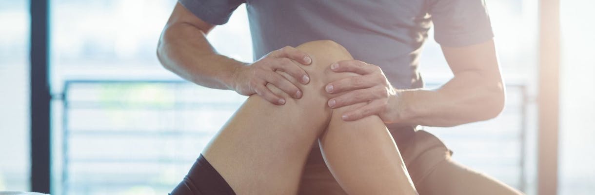
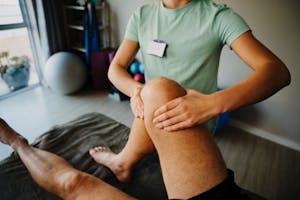
What conditions can we rehabilitate?
Manual therapy has been proven to be an important part of an overall conservative treatment plan for a variety of conditions including:
- Arthritis
- Chondromalacia
- Patellofemoral Pain
- Ligament Sprains
- Tendonitis/Bursitis
- Tendon/Muscle Strains
- IT Band Syndrome
- Post Surgical Conditions
Manual therapy is typically part of a complete treatment plan that is customized for your specific needs. Give us a call to learn how we can incorporate manual therapy into your rehabilitation program.
Physical therapy for Knee Pain
Visit our Medical Library for more information on knee pain.